Hello friends, we will learn about the Golgi complex in this article, including its definition, structure, discovery, functions, and more. So, let’s start without wasting time.
What is the Golgi complex?
Golgi complex definition –
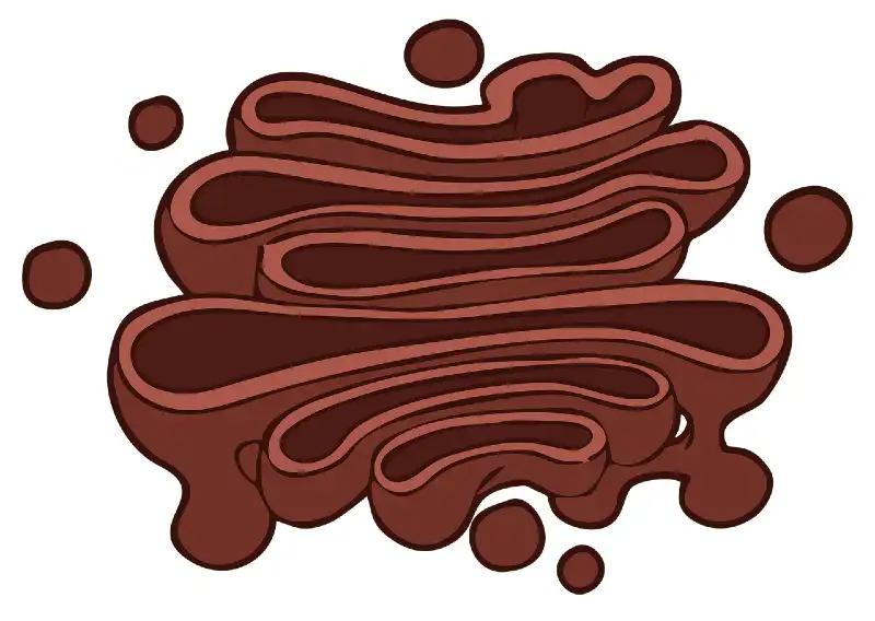
The Golgi complex is a differentiated protein of the cell’s vacuolar system. Morphologically and functionally, it is related to the endoplasmic reticulum on one side and the secretory vesicle on the other.
Who discovered the Golgi complex?
It was discovered by Camillo Golgi in 1898 in the Nerve cells by the Silver staining method. After the Discoverer’s name, it is known as the Golgi complex, Golgi apparatus, or Golgi bodies.
Where is the Occurrence of the Golgi complex?
The Golgi Complex occurs in all living cells except prokaryotic cells, R. B. Cs mature sperm, and sieve tubes in plants. In plant cells, it is called a dictyosome (Gr. dices: net).
Ultrastructure Golgi complex
Golgi Complex comprises stacks of membrane-bound space, which are of the following types :
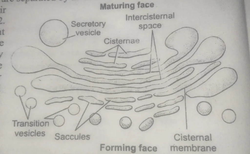
Cisternae –
Cisternae form the central plate-like Part of the Golgi complex. They consist of stacks of tubular or flattened compartments about 20 angstroms wide, enclosed by unit membranes.
These are stacked in parallel bundles, one above the other, and are separated by an intracisternal space of about 20 – 30 microns (200-300 angstrom).
The number of sternae varies from 3 to 12. They are somewhat crescent and arranged concentrically, with a convex surface toward the nuclear envelope or endoplasmic reticulum and a concave surface toward the plasma membrane.
Cisternae in a stack are arranged in a specific order. Those on the convex side are small.
This site represents the forming face of the Golgi apparatus. The transition vesicle and tubules that detach from the endoplasmic reticulum fuse here to create New cisternae.
The opposite concave face of the cisternal sack represents the maturing face. It is associated with secretory vesicles and vacuoles. These are formed by the Dilation of the ides of cisternae due to the accumulation of secretory products formed by the concentration in Golgi cisternae. These vesicles coalesce and finally create large vacuolar structures called zymogen granules or lysosomes.
Transition vesicles –
These are small drop-like structures about 400 angstroms – 800 angstroms in diameter. These are associated with the convex forming face of Golgi cisternae and lie next to the smooth endoplasmic reticulum developed from the cisternae of Endoplasmic Reticulum and fuse to form Golgi cisternae. This region represents the zone of exclusion, an area of transition from the endoplasmic reticulum to the Golgi cisternae.
Secretory vesicles –
These are present on the side and the maturing face of the Golgi. These are pinched off from the trans face of Golgi cisternae. These contain Golgi secretory products and are finally converted into zymogen granules or lysosomes.
Coated vesicles –
In some cell types, the secretory vesicles are coated with a history layer of protein clathrin. These are called coated vesicles, and they are associated with the Secretion of highly specialized cellular products.
Golgi vacuoles –
These are large, rounded presents on the maturing face of the Golgi. These are created by the expanded cisternae or by the fusion of secretory vesicles. The vacuoles are filled with some amorphous or granular substance.
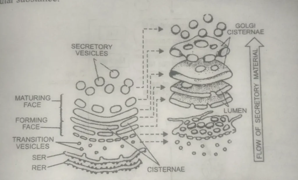
Polarity and membrane flow in Golgi –
The Golgi bodies are polarized structures. Their forming face lies close to the nuclear membrane or SER. Here, the new Golgi cisternae continue to be formed from the coalescing of transition vesicles.
On the maturing face or trans face, the cisternae break down into secretory vesicles va,cuoles, or zymogen granules. Thus, Golgi’s two faces are different.
Golgi, Endoplasmic reticulum, and lysosome association are reflected in the GERL region (Where G-represent Golgi ER-endoplasmic reticulum and L-lysosome).
The region between the Golgi and plasma membrane is where secretory vesicles are converted into zymogen granules by the concentration of products synthesized in RER.
What is the function of the Golgi complex?
The Golgi complex is vital, as it performs various functions in different types of cells. Some of them have been summarized in the following table.
Secretion –
Golgi are mainly associated with the cell’s secretory activity. Substances continuously travel from the cell to the outside through the Golgi’s channels. These substances are synthesized elsewhere in the cells, concentrated, and modified by the Golgi complex.
Finally, these are secreted outside. The Secretion Table summarizes the various substances secreted by the Golgi in different cell types. These include enzymes,e hormones, endocrine secretions, mucus, etc.
Concentration and storage of secretory product –
Golgi is associated with the concentration, storage, condensation, and Packaging of materials. The glycoprotein and lipoprotein are concentrated and packed in secretory vesicles that bud off Golgi cisternae.
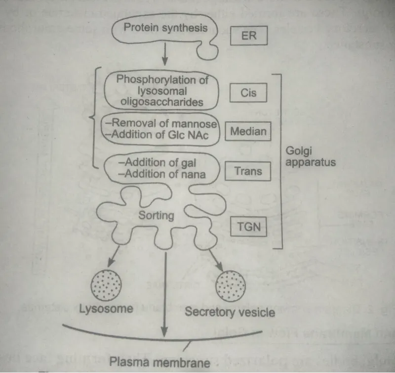
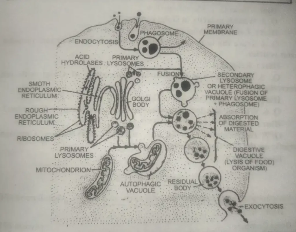
Formation of lysosome and vacuoles –
Primary lysosomes are formed from Golgi cisternae In the same way as the secretory vesicles.
Summary of functions of Golgi complex in different types of cells.
| Cell Type | Golgi Functions |
| Exocrine cells of the pancreas. | Secretion of zymogen (digestive enzymes-protease, lipase, carbohydrates, and nucleases). |
| Goblet cells of the intestinal mucosa | Secretion of mucus and zymogens. |
| Paneth cells of the intestine | Secretion of proteins. |
| Brunner’s glands of duodenum and ileum | Secretion of mucopolysaccharidoses and enzymes. |
| Hepatic cells of the liver | Transformation and secretion of lipids. |
| Follicle cells of the thyroid gland | Prothyglobulins (Hormones) |
| Plasma cells of blood | Immunoglobulins (hormones). |
| Cells of the alveolar epithelium of mammary glands. | Secretion of milk proteins. |
| Plant cells | Secretion of protein and cellulose. |
| Endothelial cells of blood vessels | Sulphation reactions. |
Synthesis of glycoproteins –
For some years, the Golgi was believed to be a passive channel for transporting materials synthesized elsewhere in the cell. However, as you know, it is now established that the Golgi facilitates linking carbohydrates and proteins to form glycoproteins.
Synthesis of carbohydrates –
The Golgi apparatus is considered to be the main agency for building large molecules of complex carbohydrates from simple sugars.
For example, in plant cells, the synthesis of protein and other carbohydrates and the formation of Polymucosaccharides occurs Inside the Golgi.
Formation of cell plate –
The Golgi Complex produces vesicles during the anaphase of Mitosis. These vesicles fuse to form the cell plate.
The pectic material and other substances necessary for forming cell walls are synthesized and secreted by the Golgi complex.
Formation of acrosome –
The acrosome of sperm develops from the Golgi complex.
Formation of acrosome or role of Golgi body in spermatogenesis
That form of spermatozoon is derived from the Golgi Complex of the spermatid.
In a spermatid, the Golgi complex comprises numerous vesicles or small vacuoles in the center, surrounded by several rows of concentrically arranged cisternae. As the differentiation proceeds, the arrangement of cisternae becomes irregular, and one or two vacuoles enlarge to replace the vesicles.
Inside each large vacuole appears a small, dense body of the acrosomal granule. The pro acrosomal granule and vacuole are formed of polysaccharides. If more than one vacuole and pro-acrosomal granules are formed, these fuse together so that only one is left.
The vacuole with its acrosomal granule enlarges in size, migrates towards the anterior pole, and gets attached to the tip of the elongated nucleus, forming a cap (cap’s stage). The pro-acrosomal granule enlarges further and forms the acrosomal granule.
It forms the core of the acrosome (acrosomal stage). The vacuole loses its liquid content and spreads over the acrosomal granule and half of the spermatozoon. It is known as the acrosome cap. The remainder of the Golgi body undergoes a gradual regression and is discarded as ‘Golgi rest‘ together with the cytoplasm of the spermatid.
Friends, if you like the information, share it as much as possible.
Thank you