If you are curious about cells and want to know about them from the beginning, then this article is for you. You will learn about the cell if you read this article until the end. So, let’s start without wasting time!
What is a Cell?
A cell is the fundamental unit of life. All living things, from microorganisms to giant organisms, are composed of cells. Without cells, there would be no life.
Examples of living things with cells:
Humans, animals, microorganisms, plants, etc.
Examples of non-living things without cells:
Pens, stones, wood, iron, etc.
Why is the cell the unit of life?
Calls are capable of independent existence and performing all the essential functions of life, such as respiration, nutrition, and excretion.
How many cells does a human have?
A human is a multicellular organism consisting of trillions of cells that work together to perform all necessary functions, such as respiration, nutrition, excretion, movement, and reproduction.
Amoeba:
A single-celled organism.
Amoeba is a unicellular organism with a single cell that performs all vital functions like nutrition, excretion, nutrition, locomotion, and reproduction. This demonstrates how a single cell can sustain life independently.
Why is the cell the structural and functional unit of life?
Cells are responsible for the structure and function of all living organisms, from humans and animals to plants.

Who discovered the cell?
Robert Hooke’s first objective cell was in 1665, but they were dead. Anton von Leeuwenhoek saw the first living cell in 1674.
Who discovered the nucleus?
Robert Brown discovered the nucleus.
What is cell theory?
In 1838, Matthias Schleiden, a German botanist, proposed that all plants are made up of different types of cells that join together to form tissues.
Shortly after, in 1839, Theodore Schwann, a British zoologist, observed a membrane surrounding animal cells. This membrane is known as the plasma membrane or cell membrane.
Their combined work led to the cell theory, which states that all organisms comprise cells and a membrane covers them. However, this theory initially did not explain how new cells arise during organism’s development.
In 1855, Rudolf Virchow completed the cell theory by proposing that all cells come from pre-existing cells through cell division.
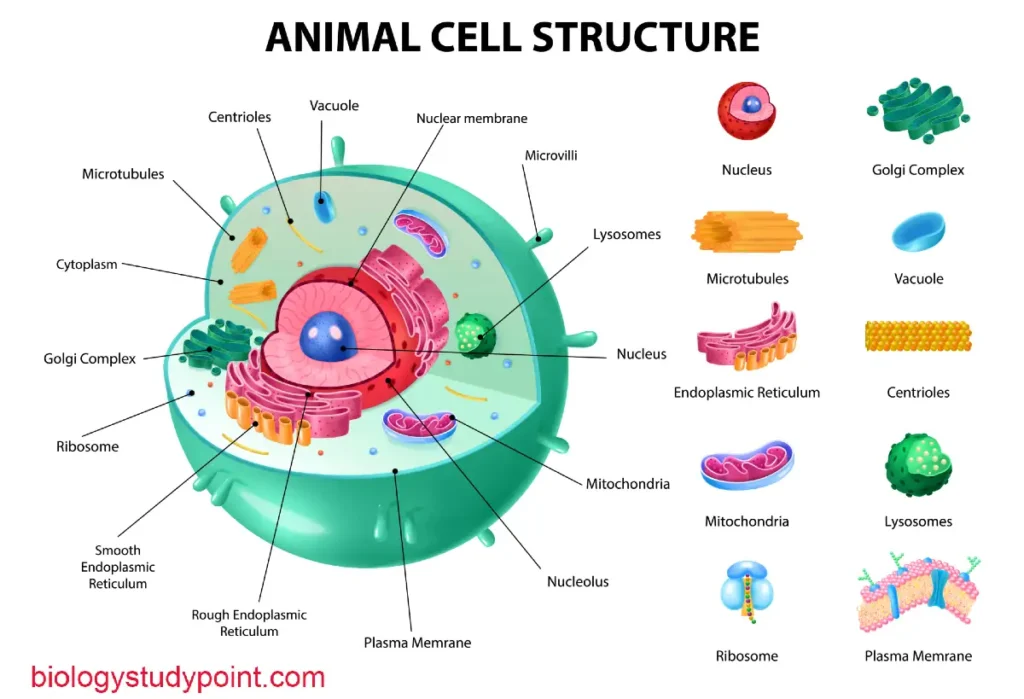
What’s inside a cell?
The main components of a cell are:
- Cell membrane: controls what enters and leaves the cell.
- Cytoplasm: A jelly-like substance that fills the cell and contains various organelles.
- Nucleus: the cell’s control center containing genetic material (DNA).
Prokaryotic vs Eukaryotic cells
There are two main types of cells.
- Prokaryotic cell: a more spartan cell that lacks a true nucleus and membrane-bound organelles. Examples include bacteria and archaea.
- Eukaryotic cell: a more complex cell with a membrane-bound nucleus and other organelles. Examples include animal, plant, and fungal cells.
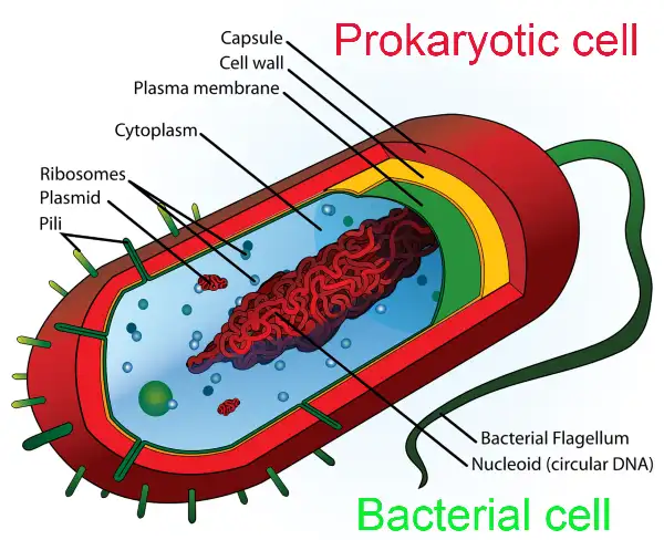
Structure of a prokaryotic cell
Prokaryotic cells have a simple structure compared to eukaryotic cells. Here are their key features:
- The cell wall (except for mycoplasma) provides rigidity and protection.
- Cell membrane: controls what enters and leaves the cell.
- Cytoplasm: contains the cell’s genetic material (DNA) in a free-floating state (not enclosed within a nucleus).
Ribosomes: produce protein (these are found in both prokaryotic and eukaryotic cells).
How many types of prokaryotic cells are there?
There are three main types of prokaryotic cells: bacteria, cyanobacteria (formerly known as blue-green algae), and mycoplasma. Mycoplasma is also placed within a group called Pleuropneumonia-like organisms (PPLO).
You may be wondering why mycoplasma, my age, is classified as a PPLO.
You might be wondering why mycoplasma is classified as a PPLO.
The reason is that bacteria and cyanobacteria have a cell wall, while mycoplasma does not. This lack of a cell wall gives mycoplasma its flexible shape. Therefore, the PPLO group includes prokaryotic cells that lack a rigid cell wall.
Prokaryotic cells are significantly smaller than eukaryotic cells.
Their more straightforward structure allows them to divide much faster than eukaryotic cells.
Which of the three types of prokaryotic cells is most common?
Bacteria are the most abundant type of prokaryotic cell. You’ve likely heard of more bacteria than the other two types because they exist in vast numbers.
Mycoplasma and the PPLO group
Mycoplasma is a type of bacteria that Lacks A cell wall, resulting in a constantly changing shape. Because of this unique characteristic, it’s classified within the pleuropneumonia-like organism (pplo) group.
Prokaryotic cell size and reproduction
Prokaryotic cells are generally much smaller than eukaryotic cells and reproduce faster through binary fission.
Types of bacteria
There are four main shapes of bacteria:
- Bacillus: rod-shaped
- Coccus: spherical
- Vibrio: Comma-shaped
- Spirillum: Spiral-shaped
Not all prokaryotic cells have a cell wall. Mycoplasma is the exception.
There’s a cell membrane following the cell wall (if present). Inside the cell membrane lies a thick, jelly-like liquid called cytoplasm.
Within this cytoplasm, the genetic material (DNA) exists in a “naked” state. A nuclear membrane does not surround it because prokaryotic cells lack a nucleus. Since there’s no nucleus, an atomic membrane wouldn’t be present.
Let’s clear up the concept of naked DNA.
Eukaryotic cells have a nucleus that houses their DNA. This DNA is about 2.2 meters long and needs to be tightly packed to fit within the nucleus. Histone proteins help with this packaging process.
Think of histone proteins as spools that DNA wraps around. If DNA is not wrapped around these spools (histones), it’s considered naked.
Therefore, naked DNA refers to prokaryotic DNA not packaged with histone proteins. This DNA is typically called genomic DNA and exists as a single circular chromosome.
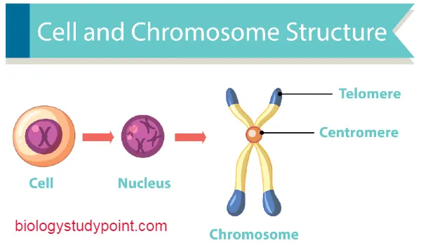
What is a chromosome?
A chromosome is a long molecule of DNA that contains genetic information. In bacteria, chromosomes are circular pieces of DNA. The two ends of the DNA molecule are joined together to form a circle.
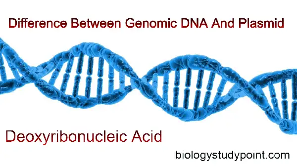
Difference between Genomic DNA and Plasmid
Genomic DNA:
This is the primary DNA molecule in a cell and contains all the essential instructions for cell function.
Plasmid:
A plasmid is a small circular piece of DNA separate from the genomic DNA. Plasmids often carry genes that provide bacteria with advantages such as antibiotic resistance.
Functions of plasmids:
Plasmid in bacteria: special DNA with unique functions
Plasmids are unique DNA molecules found in some bacteria. They come in various types and serve numerous functions. A critical function of antibiotic resistance is mediated by R-plasmid (resistance plasmids). Understanding plasmid and antibiotic resistance
Let’s break this down:
- Genes: these instructions encoded in DNA and plasmids often carry small genes.
- R-plasmids: these are plasmids that confer antibiotic resistance to bacteria. They don’t resist against whom but rather against specific antibiotics.
Example: Plasmids and Multiple Antibiotic Resistance
Imagine a bacterium with a plasmid containing two genes. One gene resists ampicillin, while the other offers resistance to tetracycline (both being antibiotics).
How does plasmid affect antibiotic treatment?
If you get sick from this bacteria, a doctor might prescribe an ampicillin resistance gene, but the antibiotic won’t be effective. The same goes for tetracycline.
However, the bacteria will likely die if the doctor prescribes streptomycin, an antibiotic the bacteria lacks resistance to (no gene on the plasmid). This highlights the importance of using the correct antibiotic to fight infections.
Mesosome: Infoldings in Prokaryotic Cells
The cell membrane can fold inwards in prokaryotic cells to create a mesosome structure. Mesosomes come in three primary forms: vesicles, tubules, and lamellae.
These infoldings are thought to play several roles in the cell, including:
- Respiration: They may aid in processes related to cellular respiration.
- DNA replication: They might be involved in facilitating DNA replication.
- Cell wall formation: they might contribute to the formation of the cell wall.
- Increased enzyme activity: The increased surface area provided by mesosomes might allow for a higher concentration of enzymes.
While the exact function of mesosomes is still being explored, they are believed to be essential structures within prokaryotic cells.
What are gram-positive and gram-negative bacteria?
The story of gram stain: a method to classify bacteria
- A Scientist named Christian Gram developed a technique for classifying bacteria known as the Gram stain.
- The process involves pressing different types of bacteria in separate beakers. First, crystal violet dye is applied to both samples, staining the bacteria.
- Next, both samples were washed with water and then with alcohol. Here, “anointing” can be replaced with “stained.”
- After washing, scientists observed that the stain remained on one type of bacteria but disappeared from the other.
- Bacteria that retained the stain were classified as gram-positive, while those that lost the stain were classified as gram-negative.
The reason behind the stain: Cell wall differences
The reason for this difference lies in the cell wall composition of the bacteria. Gram-negative bacteria have a cell wall in rich (fats). During the alcohol wash, these lipids dissolved, allowing the stain to be removed.
On the other hand, gram-positive bacteria have a cell wall with much less lipid content. This allows the crystal violet stain to remain bound to the cell wall after the alcohol wash.
Flagella: the propellers of bacteria
Not all bacteria can move. Those that can have a thin tail-like structure are called flagellum. It’s attached to the cell envelope, which consists of three main parts:
- Filament: this is the longest part of the flagellum and extends outward from the cell surface. It’s the primary structure responsible for propulsion.
- Hook: This curved structure connects the filament to the basal body.
- Basal body: embedded in the cell envelope, the basal body acts like a motor, rotating the flagellum to propel the bacterium.
It’s important to note the difference between flagella and pilli/fimbriae. While flagella are for movement, pili, and fimbriae are hair-like structures on the cell surface that help bacteria adhere to the surface or other cells. They don’t contribute to swimming speed.
Pili and Fimbriae: tiny hair-like structure with prominent roles
Pili and fimbriae are hair-like appendages found on the surface of many bacteria. While they may look similar, they have slightly different functions.
- Pili (singular: pilus): These are typically longer and less numerous than Fimbriae. Pili are made up of a Special protein called pilin. Their primary function is to help bacteria conjugate, which is transferring genetic material between two bacterial cells.
- Fimbriae (singular: Fimbriae): These are shorter and more numerous than pili. Fimbriae are also composed of proteins but not necessarily pilin. Their primary function is to help bacteria adhere to the surface, including Rock, host tissues, or other bacteria. This attachment allows bacteria to colonize a specific environment or interact with the host cell.
Also, read –
What is the animal kingdom? Complete notes.
What is the plant kingdom? Full Information.
What is biology’s definition and branches?
What is a cell wall? Complete notes
Ribosome and inclusion body in prokaryotic cells
Ribosomes are tiny structures found within prokaryotic cells responsible for protein synthesis. Unlike some cellular components, they lack a surrounding membrane. Each ribosome is roughly 15 to 20 nanometers in size and is made up of two subunits:
- A small subunit (30S) is crucial in initiating protein synthesis.
- Large subunit (50S): this subunit is responsible for protein synthesis’s elongation and termination stages.
These two subunits come together to form the complete functional 70S ribosome.
What’s inside a ribosome?
Ribosomes primarily contain two main components:
- Proteins: This protein provides structure support and helps with the various steps of protein synthesis.
- Ribosomal RNA (rRNA): this unique type of RNA molecule catalyzes protein assembly.
Polyribosome: teamwork in protein synthesis
A polyribosome, also known as a polysome, is not a single chain. Instead, its complex structure is formed when multiple ribosomes are attached to a single messenger RNA (mRNA) molecule. Imagine mRNA as a set of instructions and ribosomes as tiny factories that build protein based on those interactions.
Multiple ribosomes working on the same mRNA molecule simultaneously can increase protein production rate significantly. It’s like having numerous construction crews working simultaneously on different sections of the same building.
Which is called the protein factory.
Ribosomes are called the factory of protein because ribosomes make proteins.
Inclusion body
Some substances are not immediately used in prokaryotic cells, and these remaining substances gather in a structure in the cytoplasm. This structure is called the inclusion body.
Examples include phosphate granules, cynophycin granules, and glycogen granules.
Who is the remaining substance?
Phosphate granules, cyanophycin granules, and glycogen granules. It is found in blue-green, violet, and green photosynthetic bacteria.
So, friends, we have been studying prokaryotic cells thoroughly until now.
We will study the eukaryotic cell now, so let’s start learning.
What is a eukaryotic cell?
Those cells in which a well-developed nucleus or nuclear envelope is found are called eukaryotic cells.
In which type of organism are eukaryotic cells found?
Eukaryotic cells are found in protozoans, fungi, plants, and animals.
Both membrane-bound and membrane-less organelles are found in the cytoplasm of eukaryotic cells. Membrane-bound cell organelles include mitochondria, Golgi complex, lysosome, endoplasmic reticulum, chloroplast, and nucleus.
The membrane-less organelles are the ribosome. You have read about this in prokaryotic cells. In eukaryotic cells, the cytoplasm moves. That is, the cytoplasm moves inside. A cellular skeleton is also found in it.
Now, what is meant by a cellular skeleton? The cells of plants, fungi, and Protista have a cell wall that maintains their cell shape, but the cells of the Animalia Kingdom do not have this. So something is needed to keep their shape.
Difference between Plant cells and Animal cells
| Plant cell | Animal cell |
|---|---|
| They have a cell wall. | They do not have a cell wall. |
| Chloroplast is found in these. | Chloroplast is not found in these. |
| there is a big vacuole in them. | There is no vacoule in them. |
| They do not have a centrosome. | There is no vacuole in them. |
Microscopic fibers are found in animal cells, which form the cell skeleton and maintain its cell shape.
Now, we will talk about plant and animal cells, so let’s also study them.
Mostly, we see only plants and animals. Both of these are eukaryotic cells. Yet, there is a difference between these two cells.
Frequently Asked Questions
Which is the smallest cell?
Mycoplasma is 0.3 micrometer.
Which is the largest cell?
The largest cell is the egg of the ostrich.
Which is the longest cell?
The longest cell is a nerve cell.
Which is the smallest cell in a human?
The smallest cell in a human is the sperm.
What is the largest cell in a human?
The largest cell in an ovum.
What is the powerhouse of the cell?
Mitochondria are called the powerhouse of the cell.
What is the cell wall?
The covering of the cell, which maintains the shape of the cell, is called a cell wall. The cell wall is found in all types of cells except animal cells. It is located between the glycocalyx and the plasma membrane.
Types of cells –
There are two types of cells: prokaryotic cells and eukaryotic cells.
Who discovered the living cell?
The living cell was discovered by the scientist Anton Van Lievenhack in 1674.
Who discovered the cell and how?
Robert Hooke first discovered the cell in 1665 in the plant’s bark.
Who discovered the dead cell?
Robert Hooke discovered the dead cell.
Names of the organelles –
Mitochondria, Golgi complex, Lysosome, Endoplasmic reticulum, Chloroplast, Nucleus, and Ribosome.
The outermost layer of the animal cell.
cell membrane.
What is cytoplasm?
Inside the cell, there is a thick fluid called cytoplasm.
How many chromosomes are found in a human cell?
46 chromosomes are found. There are 23 pairs.
When was the cell discovered?
In 1665.
Where does protein synthesis take place inside the cell?
In ribosome.
What is the scientific name of a goldfish?
The scientific name of the goldfish is Carassius auratus.
What is the cell wall made of in a plant cell?
It is made of Cellulose.
Conclusion
I hope you liked the information about cells. If so, please share it with your friends so they can benefit from it, too.
Thanks