Today, we will explore a fundamental topic in biology: the differences between prokaryotic and eukaryotic cells. Let’s begin if you’re ready to dive into this fascinating subject.
Differences between prokaryotic and Eukaryotic cells.
| Feature | prokaryotic cell | Eukaryotic cell |
| Size | 1 to 10-micron meter | 10- 100 Micron meter |
| Cell wall | Noncellulose is composed of amino Sugars and muramic acid. Blue-green algae have some cellulose. | Absent in animal cells and present in plant cells consisting mainly of cellulose. |
| Capsule | Divide by simple fission, a spindle is not formed, and there is no mitosis and meiosis. | Absent |
| Cytoplasm | No streaming endocytosis or exocytosis. | Cytoplasmic streaming endocytosis and exocytosis are seen. |
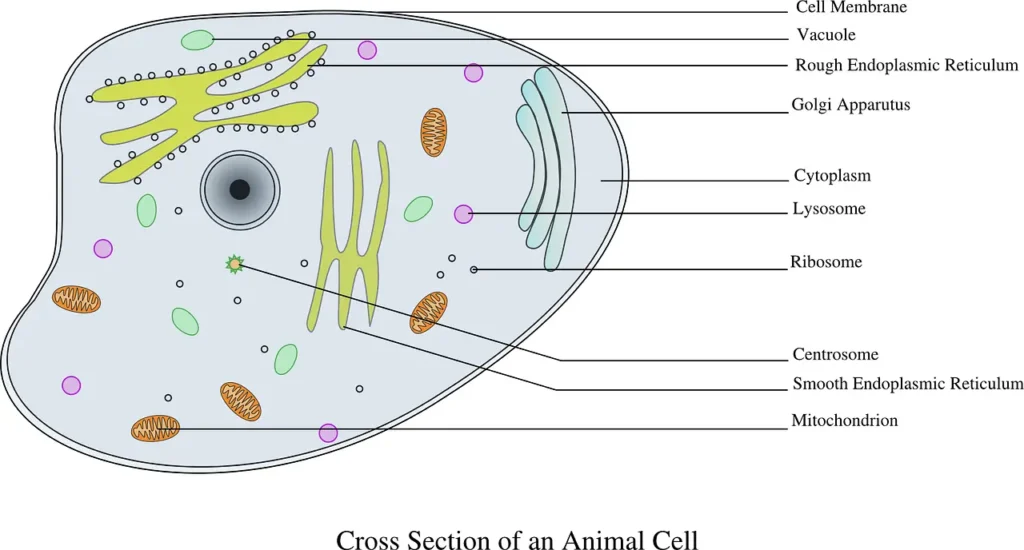
| Golgi apparatus | Absent | and concerned with cell secretion. |
| Lysosome | Absent | Plays a digestive Role. |
| Ribosome | The 70S ( 50S+30S; is Randomly scattered in the cytoplasm. | Divide by simple fission, a spindle is not formed, and there is no mitosis or meiosis. |
| Endoplasmic reticulum | Absent | Concerned with synthesis and cellular transport. |
| Microtubules and microfibrils | Absent | Present in the cytoplasmic matrix. |
| Mitochondria | Absent | Concerned with cell respiration and liberation Of energy. |
| Photosynthetic apparatus | In the form of membranes with chlorophyll-a in blue-green algae and bacteriochlorophyll in bacteria. | The 80S was found attached to the ER membrane and free in the cytoplasm. |
| Nuclear membrane | Absent | Present |
| Nucleoplasm | Not differentiated from the cytoplasm. | Separated from the cytoplasm by the nuclear membrane. |
| Nucleolus | Absent | Present |
| Vacuole | Absent | Present only in plant cells. |
| Chromosome | The single circular structure is formed of DNA only; No histone nucleus is absent. | More than one composed of chromatin (DNA and a basic protein called histones) is enclosed in the nuclear membrane to form the nucleus. |
| Flagella | Present in some species but does not have a 9 + 2 fibrillar structure. | Present in some messages has a 9 + 2 fibrillar structure. |
| Hereditary material | DNA (circular) | DNA (very long linear) |
| Cell division | They are located on the plasma membrane and are pushing. | Cells divide by mitosis or by miosis. |
| Respiratory enzymes | Transcription occurs in the nucleus, and translation occurs in the cytoplasm. | Are enclosed in mitochondria. |
| transcription and translation | Occur in the cytoplasm. | Transcription occurs in the nucleus and translation in the cytoplasm. |
| Examples | Bacteria, blue-green, algae, and mycoplasma. | Protozoa, algae, metaphyta, and metazoa. |
Differences between plant and animal cells.
Both plant and animal cells are eukaryotic. These have many features in common but differ in the following characteristics.
| Plant cell | Animal cell |
| The cell wall formed of cellulose is present outside the plasma membrane. | A cell wall is absent in all animal cells. |
| Gali complexes district is well-developed and lies near the nucleus. | Vacuoles are Absent in all typical animal cells. |
| Chloroplast is present in all green plant cells. | Chloroplast or absent. |
| Plant cells lack centrioles and centrosomes. | Animal cells possess centrioles and centrosomes. |
| Golgi complex is found scattered and is called dictyosome. | Gali complexes district and well-developed it lies near the nucleus. |
| Cytokinesis takes place through cell plate formation. | Cytokinesis occurs by constriction in the cell cytoplasm. |
What is a Bacterial cell?
Bacterial cells are typical and most common prokaryotic cells.
Structure of bacterial cell
- Size and shape –
The bacteria are non-cellular, your microscopic prokaryotic cells. The average is 1.25 microns in diameter. The smallest Bacteria are diester pneumosintes, about 0.15 T to 0.3 Micron in length, and the largest bacterium is spirillum, all volutions 13 to 15 Micron in length.
Thoracal inform (cocci) or rod-shaped (bacilli) Comma, comma-shaped spirillum, or coiled and screw-like (spirochaetes).
2. Structure –
Bacteria cells consist of the following structures:
(1) Capsule –
The number of bacteria in the slimy capsule is around the bacteria cell outside the cell wall. It is composed of polysaccharides and serves as an additional protective covering.
(2) Cell wall –
A strong, rigid cell wall covers all bacterial cells. It is about 49 to 140 microns thick and lies outside the plasma membrane. It comprises carbohydrate polypeptide peptidoglycan Thai Koi acid polyphosphate polymer, some lipids’ phosphorus inorganic salts, and a derivative called muramic acid. It also contains amino acids, such as diaminopimelic acid.
(3) Plasma membrane –
The bacterial plasma membrane is similar to the plasma membrane of eukaryotic cells. But it contained oxidative or respiratory chain enzymes. The bacterial plasma membrane is modified to form varied structures:
Mesosomes – In Tubercle bacillus and Bacillus subtilis, the plasma membrane is invaginated into whorls of convoluted membranes called mesosomes or chondroid.
These contain enzymes of the electron transport system. Mesosomes help in respiration secretion and synthesis of material for the cell walls; they receive DNA during conjugation and are sites of DNA-replication enzymes. These also help distribute chromosomes (DNA) to daughter bacterial cells.
Desmosomes: In Thiovulvum majus, The plasma membrane sinks Deep into the Cytoplasm, forming multi-layered structures called desmosomes.
(4) Cytoplasm –
The bacterial Cytoplasm contains fats, glycogen, proteins, volutin granules, etc. Certain photosynthetic bacteria have chromoplasts helping bacteriochlorophyll or other photosynthetic pigments ( carotenoids).
The pigment and the enzymes are to be associated with the internal membrane, arranged as lamellae or tubeless vesicles. These create the so-called Chromatophores.
The Cytoplasm contains 70S ribosomes. These are also called bacterial ribosomes. These occur freely in the cytoplasm or form polyribosomes. Each bacterial ribosome consists of two subunits, 30s and 50s.
(5) Nuclear material or nucleoid or genophore:
It consists of a helical DNA molecule that forms a circular chromosome stop it leads between the Santa Clause and is not enclosed in a nuclear Membrane. The clear area of the Cytoplasm, along with the bacterial chromosomes, is called nucleoid or genophore.
It is folded into several super-coiled loops.
(6) Plasmids –
E. coli and Many bacteria have many small double-stranded circular DNA molecules other than the bacterial chromosome. These are called Plasmids. These have their Individuality and replicate independently. Sometimes, plasmid DNA joins bacterial chromosomes and becomes their integral part. Such a structure is called episome.
E.coli has three types of Plasmids.
(a) F factors – These are also called sex factors. The bacterial cell having an F factor is called F+ cells, which are Donor cells. Without the F factor, they are called F- F-cells or recipient cells. F factor initiates Conjugation between F+ and F- cells.
(b) R factors – These contain genes that provide resistance to bacterial cells against antibiotics.
(c) Col factors – These factors make the bacteria secrete colicins. These are antibiotics. Colicins destroy the cells without the cool factor.
(7) Flagella –
Flagella are fine thread-like cytoplasmic structures. These come out of the cell wall and cell coat. These help bacterial cells in swimming. The bacterial flagellum is much simpler than the eukaryotic flagellum. It is without membranous covering and is formed of a single fibril about 120–150 angstroms thick. It is composed of rounded subunits of flagellin. These units are arranged in a spiral fashion.
Based on the number and arrangement of flagella, bacteria are classified into the following types:
- Atrichous – without flagella. Examples – Pasteurella.
- Monotrichous – With one flagellum only. Examples – Thiobacillus and Pseudomonas
- Lophotrichous – with many flagella, but all on one end of the bacterial cell. Examples: Spirillum solutions
- Amphitrichous – Flagella are many and present at both ends of bacterial cells. Examples – Nitrosomonas
- Peritrichous – Flagella are present all over the body.
(8) Flagella –
Some bacteria possess whip-like flagella. A bacterial flagellum comprises a single fibril about 100-200 Angstroms thick. It arises from a basal granule and is composed of a protein flagellum.
(9) Pili or fimbriae –
Some bacteria possess fine hair-like outgrowths from their surface. These are composed of helically arranged subunits of protein – pilin and are called pili. These help in the attachment.
I hope you guys know the Difference between prokaryotic and eukaryotic.
Thanks