Hello, guys. In today’s article, we will study cilia and flagella. Like – what are cilia and flagella? What is the function of cilia and flagella? We will know the answers to many such questions today, so let’s start.
What is the definition of cilia and flagella in biology?
Definition of cilia and flagella –
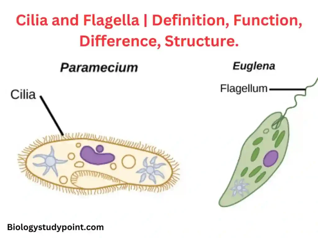
Cilia and flagella are hair-like appendages on the surface of the cells. When many hair-like appendages shorter than the cell are found on the cell’s surface, it is called cilia, and when there are 1, 2, or 4 hair-like appendages on the cell surface, they are relatively more extended than the cell. If present, they are called flagella.
Cilia and flagella are motile, hair-like appendages on the free surface of cells. Extend out of it. These cytoplasmic processes originate from the ectoplasm and extend out of it. These create water and food currents, act as sensory organs, and perform many mechanical functions for the cell.
There is no clear-cut morphological or physiological difference between cilia and flagella, except that of size. When they are few, i.e., 1,2 or 4 in number, and longer than a cell, they are called flagella, but when they are more in number and relatively short, they are known as cilia.
Also, flagella exhibit undulating motion and Beat independently, where cilia beat perpendicularly and either in metachronous (cilia of a row beating one after the other) or synchronous rhythm (all cilia of a row beating simultaneously).
What is the function of cilia and flagella?
There is no clear morphological or physical difference between the cilia and flagella, except for their shape. Both of them work the same. This Locomotion creates water and food currents, accesses sensory organs, and performs many mechanical functions for the cell.
Cilia and flagella examples
Cilia – Paramecium, respiratory tract, fallopian tube, BronchusBronchusBronchusBronchus in the lungs.
Flagella – Bacteria, sperm, euglena, etc.
What is the difference between cilia and flagella?
| Cilia | Flagella |
| Smaller than a cell (5-10µ). | Longer than the cell (150µ long). |
| Occur in large numbers (in thousands and millions). | Usually one or two per cell, but may be as many as 8. |
| Cilia beat in a coordinated fashion. Cilia of a transverse row beat synchronously (beat simultaneously) and those of a longitudinal row beat in metachronous rhythm (beat one after the other). | Flagella beat independently. |
Ultrastructure of Cilia/Cilia and flagella Structure
Describe the ultrastructure of cilium or flagellum.
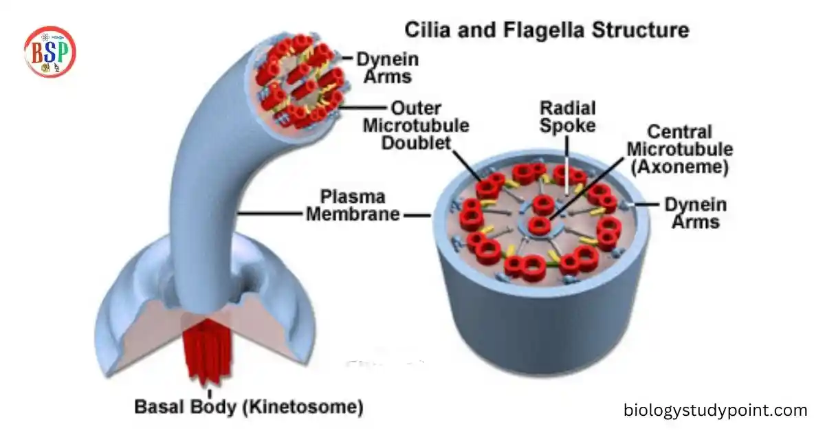
Cilia and flagella are whip-like cylindrical processes projecting from the free surface of the cell. These originate from their basal bodies embedded in the cytoplasm—the basal bodies form their kinetic centres. In size, these vary from 5µ to 10µ only. Flagella in a cell range from one to four or more and attain a size up to 150µ.
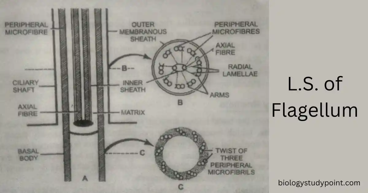
Shaft or Axial Filament –
According to the recent concept, a cilium or flagellum consists of three parts: a cylindrical shaft approximately 0.2µ in diameter with a tapering tip. This cylindrical shaft is known as the axial filament complex.
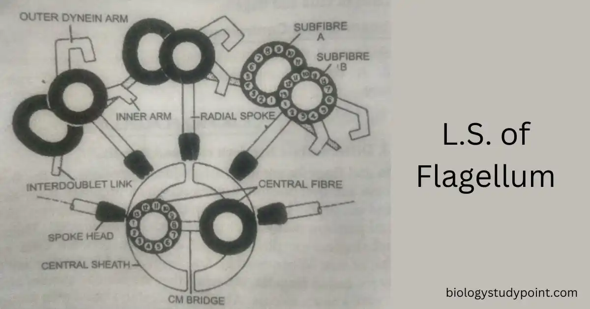
It comprises eleven longitudinal fibres, two in the centre and the other nine arranged in a cylinder around the two. The central fibres are circular in cross-section and about 200Å in diameter. Each is formed of a single microtubule, differentiated into a peripheral dense band about 50-70Å thick and a central or inner thin mass.
So, the central fibres have a tubular appearance. These are more sensitive and bear faint oblique cross-striations due to coiled fibrils within their walls. The peripheral fibres are thick, and each consists of double microtubules.
These appear as oblong cross-sections with a short microtubule or subfibre of 110-300Å and a long microtubule or subfibre of 300-350Å. The long axis is roughly parallel to the ciliary membrane. Thus, the peripheral fibres appear to be a double-barrel or figure of eight.
The two sub-fibres are unequal in size, and short projections called ‘arms‘ are seen to arise from one sub-fibre of each double fiber. The arms in each fibre are usually two, facing in the same direction on all nine fibres. The sub-fibre B is comparatively tiny and bears arms.
Its interior is denser than that of sub-fibre A. The arms are approximately rectangular structures in the longitudinal section, 50Å long and 50Å thick. The distance between the adjacent arms is about 130Å. The filaments are made up of a single type of protein, Flagella. These protein units are arranged helically.
In the distal part of the flagellum, additional fibres have also been found between the central and peripheral fibres. These are known as secondary fibrils. These are smaller but nine, each about 90Å in diameter.
A cilium or flagellum is enveloped by a continuous membrane at its base with the cell’s plasma membrane. It is about 90Å thick and has the same structure as the plasma membrane.
Trace the relationship of cilia with the basal body.
Each cilium or flagellum usually arises from a somewhat rounded shape—spherical or granular or short rod-shaped body, known as basal body.
The basal bodies of cilia are found embedded in the retractile, gelatinous ectoplasm immediately beneath the cell surface and are uniformly spaced in straight, parallel rows. The basal bodies are said to be homologous to the centriole.
In electron micrographs, the basal body appears as a hollow, cylindrical body 300–500µ long and 110–150µ in diameter with one or both ends open. It is bounded by a thick wall that encloses a central cavity filled with protoplasm of low density.
Nine groups of tubules are embedded in it. Comparable to the 9 fibres of cilia. However, each tubule comprises three units (triplets) rather than two of the cilium or flagellum. The two outer sub-fibrils of the triplet extend into the cilium or flagellum to form its doublet, while the third subfibril ends between the basal body proper and flagellum.
The association between the fibres and the basal body of a flagellum or cilium is a subject of significant variation. In mammals and protozoans, the cilia and their basal bodies are not demarcated from one another. Still, their junction is marked only by an ill-defined band of greater density, traversed by the outer fibres of the cilium, which are continuous with the similar fibres of the basal body.
In molluscs and amphibians, the peripheral fibres end into a distinct basal plate that lies above the basal body and is separated by a narrow, clear zone. In the latter case, central as well as axial fibres terminate in or near the basal plate, but in the former case, the central fibres end differently :
- In mammalian cilia, the central fibres end a short distance above the basal body into four rounded vesicles.
- Flagellates, Opalina, and Spirostomum are found in the basal granules above the basal body.
- Tetrahymena’s central fibres pass through the dense zone and extend into the basal body as a loop.
Ciliary Rootlets
Additional hair-like structures, the rootlets, extend into the cytoplasm from the basal bodies and are seen to arise; these are primarily found in the ciliated epithelial cells of mammals. Their function is presumed to anchor the basal body with the cilium.
The rootlet is found to be attached to the basal body on one end and the centrosome at the other by minute fibres. These fibres are known as rhizoplasts.
Although the basic structural plan of cilia and flagella has remained in remarkable stances, they have altered in various ways to meet special functional requirements in several instances.
Hair-like lateral processes, the mastigonemes, have been observed arising from one or both sides of the flagellum. The sperm tails represent complicated flagella. Similarly, in some instances (protozoa), the cilia fuse to form an undulating membrane. Cilia and flagella are essential in diverse physiological processes, such as Locomotion, alimentation, circulation, respiration, excretion, and sense perception.
Frequently Asked Questions
What is the short definition of cilia?
When many hair-like appendages shorter than the cell are found on the cell’s surface, it is called cilia.
What do you mean by flagella?
Flagella is a hair-like appendage found on the cell’s surface that mainly helps with the movement of the cell.
What are cilia examples?
Paramecium, Fallopian tube, Bronchus in lungs, Respiratory tract, etc.
Where is cilia?
Cilia is the present Paramecium, Fallopian tube, and Bronchus in the lungs, Respiratory tract, etc.
Where are flagella found?
Flagella are found in the sperm, euglena, bacteria, etc.
Is cilia a sperm cell?
No, cilia is not a sperm cell.
What are the two types of cilia?
Non-motile and motile are the two types of cilia.
Conclusion –
So, friends, we studied Cilia and Flagella in today’s article. Friends, if you did not like this article, please comment and explain us your suggestions and our mistakes.
Thank you so much